Age: Exposure & Diagnosis
Vulnerability of Children
In 2008, White Lake area citizens had noticed that several young people that had grown up in the area were being diagnosed with cancer.
Carpenter and Bushkin-Bedient, in Exposure to Chemicals and Radiation During Childhood and Risk for Cancer Later in Life (2013), review evidence that early-life exposure to carcinogenic chemicals and ionizing radiation results in increased risk of cancer later in life, in part because cells are rapidly dividing and organ systems are developing during childhood and adolescence. Young people have more expected years of life, during which cancers have a longer time to develop during long latency periods.
An analysis by the Environmental Protection Agency (EPA), Supplemental Guidance for Assessing Susceptibility from Early-Life Exposure to Carcinogens, concluded that cancer risks generally are higher from early-life exposure than from similar exposure durations later in life. Recognizing the unique vulnerability of children to carcinogens, the EPA recommends a 10-fold adjustment for exposures before 2 years of age, a 3-fold adjustment for exposures between 2 and 16 years of age, and no adjustment for exposures after turning 16 years of age (p. 33).
Our survey asked for all White Lake area addresses of participants, plus the year they moved in and the year they moved out of each address. With this information, I was able to look at the year each participant moved into their first White Lake area address to determine their age when they began living in the White Lake area.
Of the 1,051 participants in this project, 377 had lived in the White Lake area as children aged 0-15. Of these, 223 had been born in or moved to the area before their second birthday, and 154 moved to the area between their second and sixteenth birthdays.
Where had the Participants Lived as Children, and When?
I wanted to find out where the 377 participants had lived in the White Lake area as children, and when they had lived there. I divided them into three groups because it was important to separate full-time from part-time residents at diagnosis and to identify how many of these participants’ cancers were likely to have been counted in local cancer incidence statistics versus those that were likely to have been counted elsewhere.
These 377 participants had been diagnosed with 410 cancers. For the following analysis I used only the address at which they were living during the year that they were diagnosed with their first cancer.
Of the three groups represented on the map below, only 222 first cancer diagnoses (the 210 cancers of the blue group, and 12 of the 33 cancers of the brown group) – 59% – would have been counted in local cancer statistics, while 155 first cancer diagnoses (134 of the orange group and 21 of the brown group) – 41% – would have been counted elsewhere.
There likely was no record that many of these participants had spent all or part of their childhoods in an area that had experienced industrial pollution. This underscores the need for additional methods of data collection such as storage of Residence Histories and Exposure Histories in electronic medical records, as well as longitudinal studies and population level case-control studies (See “The Hazards/Cancer Puzzle” at the end of this report.)
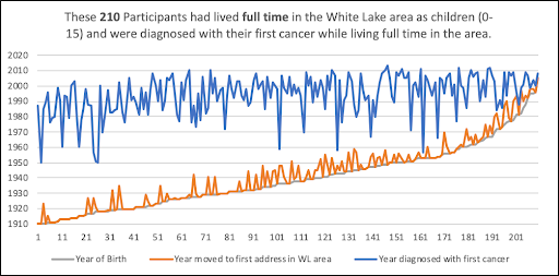
- Blue* = 210 participants that had lived full time in the White Lake area as children and were diagnosed with their first cancer while living full time in the area. These cancers would have been counted in local cancer incidence statistics.
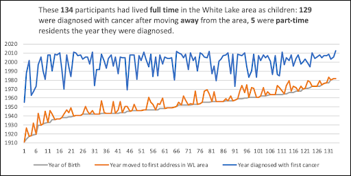
- Orange* = 134 participants that had lived full time in the White Lake area as children, but 129 had moved away prior to diagnosis and five were part-time residents at diagnosis. Their cancers were likely counted elsewhere.
- Brown* = 33 participants that had lived part time in the White Lake area as children: 12 had become full-time residents prior to diagnosis (and their cancers likely were counted in local cancer incidence data), 10 continued to be part-time residents at diagnosis, and 11 had moved away before diagnosis so their 21 cancers were likely to have been counted elsewhere.
*All addresses and year moved in were based on the child’s first address in the White Lake area except for two instances in which the description of the first residence was so vague as to not allow an approximate location and the child had lived there only briefly. For each of these, the child’s second address was used.
In many instances, two or more participants had lived at the same address during their childhood years with family members who were also participants, so the points that are viewed on the map will appear to be fewer than the number of participants represented.
First Full-Time and Part-Time Addresses of Participants that had Lived in the White Lake area as Children Aged 0-15

Map by Rick Sadler, Associate Professor, Division of Public Health, Michigan State University
The above map does not show what years the participants had lived at the indicated residences. Of the 377 participants represented on the map, 208 had been born in or moved to the area prior to 1950, the decade when the major chemical companies came to town and commenced three to four decades of pollution. Environmental hazards prior to 1950 may have been limited to those related to the tannery, located at the shoreline on the southeast end of the lake. Whitehall Leather Company stopped using bark for tanning and began using chromium in 1940.
It is interesting to note that of the 377 first cancers of participants that were born in or lived in the White Lake area as children, half (189) were diagnosed between 1950 and 1999 (49 years), and the other half (188) of the cancers were diagnosed between 2000 and 2013 (13 years). Beyond the speculation that this apparent increase in cancers could have been related to toxic exposures that these participants had experienced as children, reasons for this recent increase in numbers may include factors such as improved cancer screening and earlier diagnosis for participants in more recent years, and the possibility that more people who were recently diagnosed heard about in this project and chose to participate.
Yet the graphs below might also be a reflection of the already existing concerns of White Lake area citizens that they and their neighbors were being diagnosed with too many cancers, which then led to the launch of this cancer mapping project.
210 Full-Time Residents as Children and at Diagnosis
Year of Birth, Year Moved to White Lake Area, Year of Diagnosis with their first cancer

134 Full-Time Residents as Children but Moved Away, or Part Time at Diagnosis
Year of Birth, Year Moved to White Lake Area, Year of Diagnosis with their first cancer

33 Part-Time Residents as Children: Away, Part Time, and Full Time at Diagnosis
Year of Birth, Year Moved to White Lake Area, Year of Diagnosis with their first cancer>

Median Age at Diagnosis
White Lake area citizens were concerned that people in our community seemed to be diagnosed with cancer at younger ages than would be typical for their type of cancer. I wanted to know whether our participants had been diagnosed at younger ages when compared with statistics from the U.S. population.
Median Age at Diagnosis Table
For each cancer type below, the reference median age at diagnosis for the U.S. population is shown in the white column and the median age of our participants in the blue column.
After looking at these data, I wondered whether the 377 participants that had lived in the White Lake area as children had been diagnosed with their cancers at younger ages than those that had moved to the area at age 16 or older. Then I decided to compare median age at diagnosis of those that had lived in the area as children prior to 1950, before the chemical industries began to arrive, with those who moved to the area after 1950 and lived in the area as children while the industries were in operation.
The solid yellow columns hold the median age at diagnosis for our participants who had been born in or moved to the area as children aged 0-15, and the green columns represent those who had moved to the area at the age of 16 or older.
The yellow and green groups are each further divided to compare median age at diagnosis of participants who were born in or moved to the White Lake area prior to 1950 (before the chemical industries had come to town) with those who had moved to the area after January 1, 1950.
Median ages that are younger than the reference median age reported for the U.S. population for that cancer type appear in red print.
The total number of cancers in the group from which the median age was calculated are listed in the cells below each median age. Caution is recommended when looking at these data due to the low numbers of participants in each group, with numbers as low as one participant in several of the breakdown groups. Such small groups don’t allow for strong median calculations to occur.
As in other tables in this report, I will not try to interpret these data, other than to observe the following:
- According to comparisons using the sources found, White Lake Area Cancer Mapping Project adult participants’ median ages at diagnosis were 1-13 years younger than the U.S. reference median ages for the following cancer types: Bladder, Brain & Nervous System, Breast, Colorectal, Gynecologic, Head & Neck, Kidney & Ureter, Leukemia, Lung, Hodgkin Lymphoma, Non-Hodgkin Lymphoma, Melanoma, Mesothelioma, Multiple Myeloma, Sarcoma, Testicular, Thyroid, and Upper Gastrointestinal.
- Participants’ cancers were diagnosed at ages that were the same as or older than the U.S. reference ages for the following cancer types: Liver & Bile Duct, Pancreas & Gallbladder, and Prostate.
| Median Age at Diagnosis: Adult (age 20+) CMP cancers, compared to U.S. | Born in or Moved to WL area as child age 0-15 | Moved to WL area age 16 or older |
|||||||
| Cancer Type | Median Age at Diagnosis 1 | U.S. Popu- lation |
CMP | All: 0-15 | To WL before 1950 | To WL after 1950 | All: 16+ | To WL before 1950 |
To WL after 1950 |
| Bladder | Median Age at Diagnosis | 73 | 69 | 65 | 69.5 | 53 | 69 | 80 | 69 |
| Number Cancers in Group | 41 | 11 | 10 | 1 | 30 | 3 | 27 | ||
| Brain & Nervous System | Median Age at Diagnosis | 59 | 53.5 | 38.5 | 47 | 35.5 | 58.5 | 73 | 58 |
| Number Cancers in Group | 32 | 10 | 2 | 8 | 22 | 1 | 21 | ||
| Breast | Median Age at Diagnosis | 62 | 54 | 50 | 57.5 | 44.5 | 56.5 | 59 | 55.5 |
| Number Cancers in Group | 218 | 90 | 44 | 46 | 128 | 14 | 114 | ||
| Colorectal | Median Age at Diagnosis | 67 | 66 | 67 | 70 | 40 | 65 | 61 | 65 |
| Number Cancers in Group | 96 | 30 | 21 | 9 | 66 | 12 | 54 | ||
| Gynecologic 2 (Ovary & Uterus) |
Median Age at Diagnosis | 63 | 58 | 57 | 61 | 47.5 | 59 | 65 | 58 |
| Number Cancers in Group | 50 | 19 | 13 | 6 | 31 | 7 | 24 | ||
| Gynecologic 2 (Cervix) |
Median Age at Diagnosis | 50 | 43.5 | 38.5 | 47.5 | 33.5 | 44.5 | 84 | 43 |
| Number Cancers in Group | 16 | 4 | 2 | 2 | 12 | 1 | 11 | ||
| Head & Neck | Median Age at Diagnosis | 63-65 | 58 | 52 | 68 | 48 | 61 | 66.5 | 61 |
| Number Cancers in Group | 36 | 16 | 7 | 9 | 20 | 2 | 18 | ||
| Kidney & Ureter | Median Age at Diagnosis | 64 | 63.5 | 72 | 72 | NA | 62 | 70 | 58 |
| Number Cancers in Group | 22 | 5 | 5 | 0 | 17 | 3 | 14 | ||
| Leukemia 3 (adult, age 20+) |
Median Age at Diagnosis | 67 | 62 | 50.5 | 54 | 48 | 70 | 79 | 63 |
| Number Cancers in Group | 31 | 10 | 3 | 7 | 21 | 6 | 15 | ||
| Liver & Bile Duct | Median Age at Diagnosis | 65 | 65 | 73.5 | 73.5 | NA | 61 | 65 | 60.5 |
| Number Cancers in Group | 17 | 6 | 6 | 0 | 11 | 3 | 8 | ||
| Lung | Median Age at Diagnosis | 71 | 64 | 63 | 64 | 58.5 | 65 | 64 | 66 |
| Number Cancers in Group | 172 | 49 | 37 | 12 | 123 | 17 | 106 | ||
| Lymphoma Hodgkin | Median Age at Diagnosis | 39.5 | 35 | 33 | 47 | 22.5 | 47.5 | NA | 47.5 |
| Number Cancers in Group | 13 | 11 | 5 | 6 | 2 | 0 | 2 | ||
| Lymphoma Non-Hodgkin 4 |
Median Age at Diagnosis | 67 | 61 | 50.5 | 63 | 47 | 68 | 78 | 67 |
| Number Cancers in Group | 48 | 18 | 7 | 11 | 30 | 3 | 27 | ||
| Melanoma | Median Age at Diagnosis | 65 | 56.5 | 41.5 | 65 | 32 | 59 | 61 | 59 |
| Number Cancers in Group | 36 | 16 | 7 | 9 | 20 | 2 | 18 | ||
| Mesothelioma | Median Age at Diagnosis | 72 | 67 | 64 | 64 | NA | 68.5 | NA | 68.5 |
| Number Cancers in Group | 7 | 1 | 1 | 0 | 6 | 0 | 6 | ||
| Multiple Myeloma | Median Age at Diagnosis | 69 | 67 | 56 | 53 | 60 | 72 | 57 | 72.5 |
| Number Cancers in Group | 23 | 5 | 4 | 1 | 17 | 3 | 14 | ||
| Pancreas & Gallbladder | Median Age at Diagnosis | 70-72 | 71 | 71 | 72 | 39.5 | 71 | 76.5 | 69 |
| Number Cancers in Group | 31 | 8 | 6 | 2 | 23 | 2 | 21 | ||
| Prostate | Median Age at Diagnosis | 66 | 68 | 66 | 67 | 55.5 | 68 | 72 | 67 |
| Number Cancers in Group | 94 | 27 | 25 | 2 | 67 | 10 | 57 | ||
| Sarcoma (Bone & Joint) |
Median Age at Diagnosis | 45 | 32 | 21 | NA | 21 | 66 | NA | 66 |
| Number Cancers in Group | 7 | 3 | 0 | 3 | 4 | 0 | 4 | ||
| Sarcoma 5 (Soft Tissue) |
Median Age at Diagnosis | 61 | 51 | 45 | 60 | 44 | 72.5 | 71 | 74 |
| Number Cancers in Group | 18 | 8 | 3 | 5 | 10 | 3 | 7 | ||
| Testicular | Median Age at Diagnosis | 33 | 32 | 30.5 | 32 | 29 | 44 | NA | 44 |
| Number Cancers in Group | 10 | 6 | 1 | 5 | 4 | 0 | 4 | ||
| Thyroid | Median Age at Diagnosis | 51 | 47 | 51 | 63 | 37 | 44 | NA | 44 |
| Number Cancers in Group | 10 | 8 | 5 | 3 | 2 | 0 | 2 | ||
| Upper Gastro- intestinal |
Median Age at Diagnosis | 66-68 | 63.5 | 56.5 | 57.5 | 40 | 66 | 60 | 68 |
| Number Cancers in Group | 30 | 8 | 6 | 2 | 22 | 3 | 19 | ||
- The median ages at diagnosis should be interpreted cautiously, due to unknown or confounding variables that include:
-
- The number of participants is small, making median age calculations unreliable.
- Our cancers were diagnosed over a wide range of years, whereas the data used for SEER’s calculations appear to span a limited range of four recent years, approximately from 2013-2017.
- Some cancers, including breast cancer, may be diagnosed at younger ages in more recent years due to screening that wouldn’t have been available in the past.
- More young people than older people with cancer may have decided to participate in this cancer mapping project during the survey period.
- I was likely to have made errors in organizing cancer types into groups. Some rare cancers included within the participants’ cancer groups may not have been included in the SEER or ASCO categories that were used to calculate median age at diagnosis.
- Median age for ovarian and uterine cancers is 63. Median age for cervical cancers is 50, so ovarian and uterine gynecological cancers were listed together here, with cervical cancers listed separately. Two participants had both uterine and cervical cancers, so their two cancers appeared in each of their respective groups.
- SEER reports that the median age at diagnosis for all leukemias combined is 67, yet SEER also reports a median age at diagnosis for each of the four main types of leukemia: ALL (17), AML (68), CLL (70) and CML (65). Of the 31 White Lake area adult leukemias reported here, 7 were leukemia nos, 4 acute leukemia nos, 1 acute lymphocytic (ALL), 5 acute myeloid (AML), 7 chronic lymphocytic (CLL), 4 chronic myeloid (CML), 2 hairy cell (HCL), and 1 large granular lymphocytic leukemia (LGL).
- Thirteen survey respondents reported only Lymphoma, not specifying Hodgkin (HL) or Non-Hodgkin (NHL) lymphoma. These were presumed to be NHL.
- SEER Stat includes Ewing Sarcoma under the link, “More about this cancer” in both the “Bone & Joint” and “Soft Tissue Sarcomas” cancer groups. Four of our adult participants had this rare cancer: three were bone cancers (listed in the 0-15, post-1950 bone cancer group) and one was a soft tissue cancer (listed in the 0-15, post-1950 soft tissue cancer group).

